Tooth extractions are among the most common procedures in dentistry, performed for reasons ranging from advanced decay and infection to orthodontic needs or impacted wisdom teeth.
Patients frequently express curiosity about the physical appearance of the tooth once it has been successfully removed from its socket in the jaw.
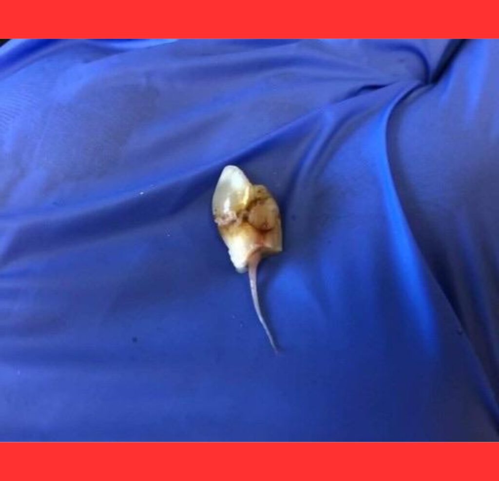
Upon extraction, the first thing noticeable is the crown of the tooth.
This is the portion that was previously visible above the gum line, protected by a layer of durable enamel.
Depending on the condition that led to the extraction, the crown may display discoloration, cavities, or structural damage.
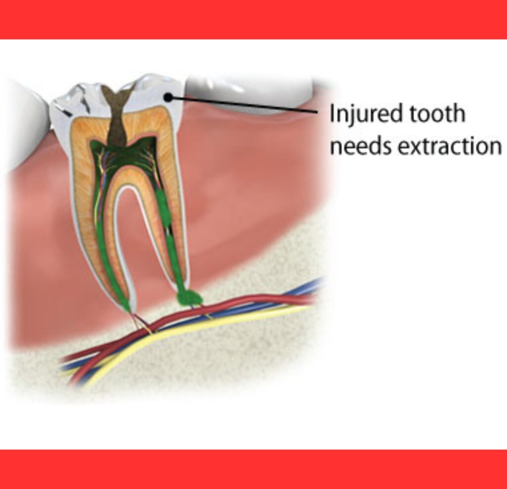
Extending from the crown are the roots of the tooth, which are embedded deep within the alveolar bone of the jaw.
These roots, often numbering one to three or more, provide the necessary anchorage and can vary greatly in length and curvature based on the tooth type.
The roots are typically a pale yellow or ivory color and are covered by a thin layer called cementum.
In a clean extraction, the entire root structure comes out intact, revealing the complex morphology that was hidden beneath the gums.
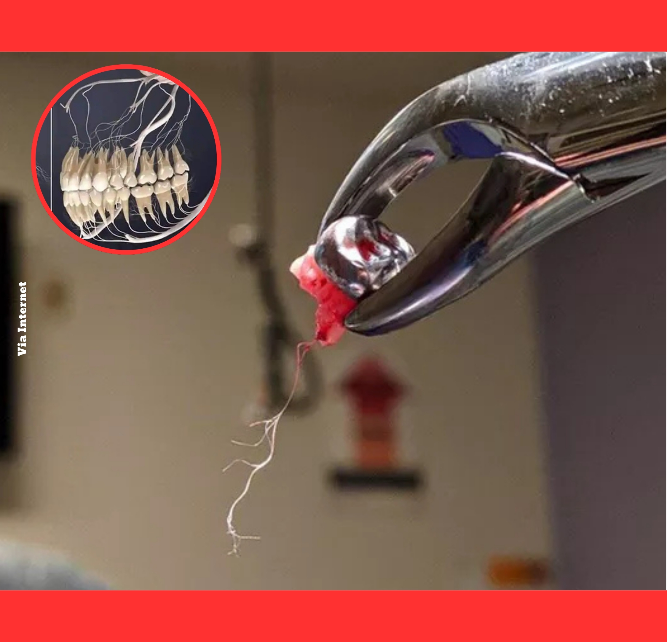
Perhaps the most intriguing element visible in some extractions is the dental pulp, frequently referred to by patients as the nerve root.
This living tissue occupies the pulp chamber in the crown and extends through narrow canals into each root.
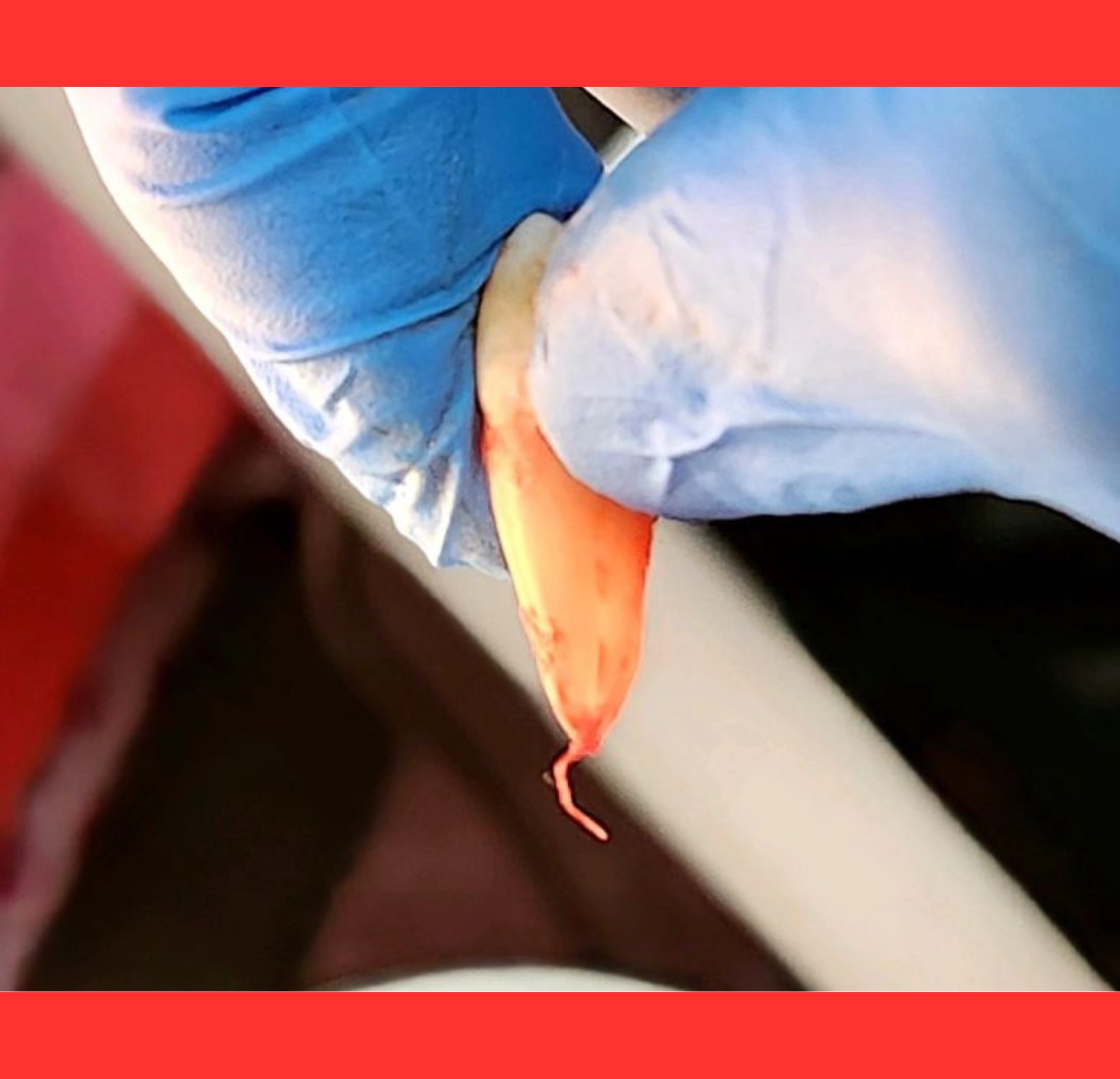
When the pulp remains attached after removal, it appears as a delicate, thread-like or stringy structure protruding from the bottom of the root.
Composed of nerves, blood vessels, and connective tissue, it once nourished the tooth and transmitted sensory signals.
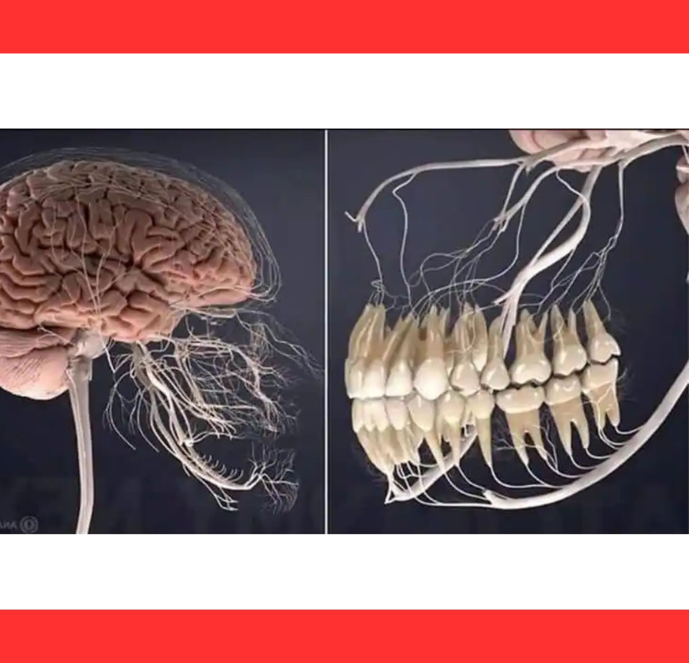
Observing an extracted tooth with its nerve root intact offers a rare educational glimpse into oral anatomy.
It underscores how teeth are not just hard shells but dynamic, living organs connected to the body’s nervous system.
Dental professionals utilize specialized instruments like elevators and forceps to gently loosen and remove the tooth while minimizing trauma.
The occasional intact nerve attachment occurs when the pulp does not fragment during the pulling process.
Following the extraction, the tooth socket is thoroughly cleaned and may be sutured if necessary.
Healing begins immediately with the formation of a blood clot, which is essential for proper recovery and preventing issues such as infection or dry socket.
Gaining knowledge about what an extracted tooth looks like, including its roots and attached nerve, can alleviate anxiety surrounding dental visits.
Maintaining excellent oral hygiene remains the best strategy to preserve natural teeth and avoid the need for extractions.